Recognize the Keys to Coding Diagnostic VATS Procedures
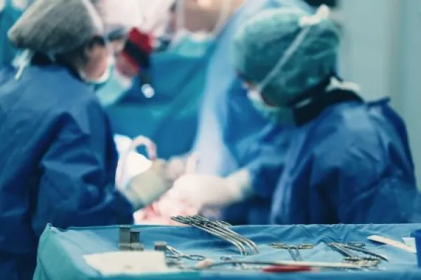
Does the provider have to perform a biopsy? Find out. During a video-assisted thoracic surgery (VATS) procedure, the physician uses a camera on a thoracoscope to visualize the inside of the patient’s chest on a video monitor. This instrument allows the physician to examine body structures, collect tissue samples, and perform other tasks with minimal invasion to the patient. Attendees learned about the surgeries and CPT® codes tied to VATS procedures in the AAPC HEALTHCON 2026 session, “Through the Scope: Coding and Clinical Insight Into VATS Lung Procedures,” compiled by Amanda Reikowsky, CMCPS, CPC, CRC, CPMA, CDEO, independent consultant with CodeWise Solutions LLC and presented by Sandy Giangreco Brown, MHA, BS, NREMT, RHIT, CCS, CCS-P, CHC, CPC, COC, CHRI, CPCO, CPC-I, COBGC, PCS, AAPC Approved Instructor, vice president of revenue integrity and education at Spire Orthopedic Partners. Keep reading to learn about diagnostic VATS procedures and how to properly code them. Get to Know VATS Surgical thoracoscopy, also known as VATS, is a procedure where the provider uses a thoracoscope with a camera to examine the patient’s chest cavity. A video monitor in the operating room displays what the camera sees as the surgeon guides it through the patient’s body to evaluate body structures. Benefits of the procedure over an open thoracotomy include smaller incisions to allow instruments and equipment to enter the thoracic cavity. These minimal incisions help reduce the patient’s pain during recovery, lower the amount of time spent in the hospital, and decrease the risk of morbidity and mortality. Recognize Diagnostic VATS Procedure Codes VATS procedures fall under diagnostic or therapeutic categories. A provider will perform a diagnostic VATS to examine the pleural cavity and lungs to evaluate possible pulmonary lesions or nodules, or if abnormalities were found on imaging exams. VATS turns into a therapeutic procedure when the surgeon determines there is a need to treat the patient. “Typically, the diagnostic procedures are going to be more sampling or looking around, and we know if the provider does a therapeutic procedure, that looking around is always included,” Giangreco Brown explained. Assign 32601 (Thoracoscopy, diagnostic (separate procedure); lungs, pericardial sac, mediastinal or pleural space, without biopsy) when the physician performs diagnostic VATS without a biopsy. This code applies to a diagnostic examination of the lungs, pericardial sac, mediastinal space, or the pleural space. “This one is basically just going in. If they’re just going in there to look around, maybe they’re going in there to try to figure out what’s going on with this patient, and they’re not doing a biopsy,” Giangreco Brown said. When the physician captures a biopsy during a diagnostic VATS procedure, you’ll assign one of the following codes: The provider might choose to collect a biopsy following a prior procedure, such as a bronchoscopy where the physician found an abnormality and now, they’re re-entering the space to do a biopsy. Simultaneously, the doctor might decide during the diagnostic VATS to do a biopsy so they can determine the next treatment steps. Dive Into VATS With Diagnostic Biopsies Use 32607 (Thoracoscopy; with diagnostic biopsy(ies) of lung infiltrate(s) (eg, wedge, incisional), unilateral) to report a VATS procedure where the physician performs a diagnostic biopsy of lung infiltrates. Infiltrates are usually abnormal substances such as pus, fluid, blood, or immune cells that fill the tissues and air sacs of the lungs. The provider uses instruments to remove a small piece of the lung for examination. If the surgeon performs a diagnostic biopsy or biopsies of masses or nodules in the lung, you’ll assign 32608 (Thoracoscopy; with diagnostic biopsy(ies) of lung nodule(s) or mass(es) (eg, wedge, incisional), unilateral). Codes 32607 and 32608 are unilateral, which means that regardless of whether the provider collects one or multiple biopsies from one lung, you’ll report each code once per lung. “If the provider is performing the procedure on the other lung, then we would want to report that as well,” Giangreco Brown explained. Example: A patient presents for VATS with diagnostic biopsies of lung nodules after chest X-rays revealed abnormalities in both lungs. The physician preps the patient, performs VATS on the right lung, and incises a small piece of the lung for examination. After the sample is collected, the provider turns their attention to the left lung. This time, the surgeon performs a wedge biopsy to examine the tissue. In this example, you’ll assign 32608 appended with modifier RT (Right side) to report the incisional diagnostic biopsy performed on the right lung. You’ll also use another 32608 appended with modifier LT (Left side) to report the wedge biopsy on the left lung. Pinpoint the Pleural Biopsy Code The pleura is the membrane lining the chest cavity and surrounding the lungs. The pleura consists of several different layers, such as: “The parietal pleura, the outer layer of the pleura, can attach itself to the chest wall, and that is where sometimes you’ll hear of patients getting pleuritis, which is an inflammation in there. The patient might present to the office saying, ‘Every time I take a breath, it hurts — it literally hurts to breathe,’” Giangreco Brown said. When the physician performs VATS and collects one or more tissue samples from the pleura, you’ll assign 32609 (Thoracoscopy; with biopsy(ies) of pleura) for the procedure. Takeaways Coding diagnostic VATS procedures correctly requires careful examination of the documentation and the CPT® guidelines. Remembering these helpful tips can take your procedure coding to the next level: Mike Shaughnessy, BA, CPC, Production Editor, AAPC