Learn Why Radiologists Use Dual-Energy CT to Evaluate Patients
Plus: You’ll be surprised by how to code DECT procedures. Dual-energy computed tomography (DECT) is an advanced CT imaging technique that acquires images using two different X-ray energy spectra, providing information about tissue composition beyond that available with conventional CT. By comparing how materials attenuate low- and high-energy X-ray beams, DECT can differentiate substances based on their atomic composition, allowing improved tissue characterization and enhancing diagnostic confidence across numerous clinical applications. Keep reading to learn more about DECT procedures and how to code the imaging studies. Get to Know DECT Although the concept of DECT was first introduced in the early 1970s, widespread clinical adoption did not occur until the mid-2000s, when manufacturers developed commercially practical scanner designs capable of incorporating dual-energy technology into routine imaging workflows. Unlike conventional CT, which acquires data using a single energy spectrum, DECT obtains image datasets at two different energy levels, either simultaneously or in rapid succession depending on scanner design. Because materials demonstrate unique attenuation characteristics at different X-ray energies, specialized reconstruction algorithms can distinguish substances such as iodine, calcium, uric acid, and soft tissue with greater specificity than conventional CT. These capabilities support improved diagnosis, lesion characterization, artifact reduction, and functional imaging without requiring additional invasive procedures. Take These Coding Considerations Into Account From a coding perspective, DECT does not have separate CPT® codes. Code selection follows the same guidelines used for conventional CT examinations and is determined by these factors: The use of dual-energy acquisition or post-processing does not independently affect CPT® code assignment or reimbursement. For example, if the provider performs a DECT study with contrast to examine the patient’s lungs, then you’d assign 71260 (Computed tomography, thorax, diagnostic; with contrast material(s)) for the procedure. Medicare and other third-party payers generally consider coverage based on the medical necessity of the underlying CT examination and applicable coverage policies rather than the imaging technology itself. Documentation supporting the medical necessity of the examination remains essential regardless of whether dual-energy technology is used. Dive Into Gout Detection Gout is an inflammatory arthropathy caused by monosodium urate (MSU) crystal deposits within joints and surrounding soft tissues. DECT has become an important noninvasive imaging tool for evaluating suspected gout because it can directly identify, characterize, and quantify urate crystal deposits. DECT is particularly valuable when clinical presentation is atypical or joint aspiration is inconclusive or impractical. Early identification of urate deposits allows prompt therapy initiation and may reduce unnecessary invasive procedures while helping prevent long-term complications such as chronic joint destruction, tophaceous disease, tendon injury, and functional impairment. Examples of chronic gout codes listed in the ICD-10-CM code book include: Because gout is associated with significant morbidity and is frequently accompanied by cardiovascular and renal comorbidities, accurate diagnosis coding can improve both patient management and long-term outcomes. Learn How Physicians Use DECT for Pulmonary Imaging DECT has expanded the capabilities of pulmonary imaging by providing both anatomic and functional information from a single examination. Material decomposition techniques generate iodine maps, perfused blood volume (PBV) images, and virtual monoenergetic images (VMI), each contributing additional diagnostic information beyond conventional CT. Map Blood Flow Through the Pulmonary System Iodine maps evaluate pulmonary perfusion by displaying the distribution of iodinated contrast material within the lungs. These maps assist in assessing regional blood flow and can identify perfusion abnormalities associated with pulmonary embolism, chronic thromboembolic disease, and other vascular disorders. Material decomposition also permits creation of perfusion maps without requiring additional noncontrast acquisition. Image Perfused Blood Volume PBV images demonstrate the distribution of blood flow throughout the pulmonary parenchyma. In patients with pulmonary embolism, PBV imaging allows simultaneous visualization of emboli within the pulmonary arteries and the corresponding perfusion defects within the affected lung tissue. PBV findings may also help estimate the physiologic severity of pulmonary embolic disease. Visualize Lesions and Vascular Structures Virtual monoenergetic images (VMI) are reconstructed from dual-energy datasets to simulate images acquired at a single photon energy level measured in kiloelectron volts (keV). Lower-keV reconstructions increase iodine attenuation, improving enhancement of contrast-filled vessels and lesions; while higher-keV reconstructions reduce beam-hardening artifacts caused by dense contrast material or metallic implants. Optimization of keV settings can improve lesion conspicuity, vascular visualization, and overall image quality. Learn How Physicians Use VMIs and VNC Images With DECT DECT is an important tool in vascular imaging by improving visualization of blood vessels while simultaneously reducing image artifacts. Material decomposition techniques generate VMIs, virtual noncontrast (VNC) images, and iodine maps that provide additional diagnostic information without requiring separate imaging acquisitions. Iodine maps assist in evaluating tissue perfusion, identifying vascular occlusions, characterizing vessel wall abnormalities, and assessing enhancement patterns of tumors and organs. Low-keV VMIs enhance iodine attenuation, making subtle vascular enhancement and minimally enhancing lesions more conspicuous. These reconstructions improve visualization of small vessels, coronary arteries, endovascular stents, and myocardial perfusion while increasing diagnostic confidence. Higher-keV VMIs reduce beam-hardening and blooming artifacts associated with metallic implants, surgical clips, coils, dense contrast material, and heavily calcified atherosclerotic plaque. Artifact reduction improves evaluation of adjacent vascular structures and surrounding soft tissues. VNC images digitally subtract iodine from contrast-enhanced examinations, reducing the need for additional true noncontrast acquisitions in selected clinical scenarios. VNC imaging has demonstrated value in evaluating endoleaks following endovascular aneurysm repair (EVAR), characterizing portal vein thrombus, and assessing various abdominal and vascular abnormalities. However, VNC images do not completely replace true noncontrast (TNC) examinations in every clinical situation and dedicated noncontrast imaging remains necessary when clinically indicated. Remember These Documentation Tips Accurate physician documentation supports appropriate coding, reimbursement, and medical necessity regardless of whether dual-energy technology is used. Documentation should include the following elements: Although documentation of dual-energy post-processing supports the clinical record, it does not change CPT® code selection. Don’t Miss These Frequently Misunderstood Concepts Misconception 1: DECT always doubles radiation exposure. Modern DECT systems generally achieve radiation doses comparable to conventional CT examinations through advances in detector technology, automated exposure control, and optimized acquisition protocols. Radiation exposure depends primarily on the clinical protocol rather than the use of dual-energy technology itself. Misconception 2: DECT requires different CPT® coding. The CPT® code book does not contain separate CPT® codes for DECT. Coding is based on the anatomical region examined and the use of contrast, which follows the same guidelines applied to conventional CT examinations. Misconception 3: Material decomposition is always perfect. Material decomposition relies on mathematical algorithms and predefined material models, typically including iodine, calcium, water, and uric acid. Image quality may be affected by severe metallic artifacts, patient motion, or materials not included within the reconstruction model. Misconception 4: VNC images completely replace TNC scans. Although VNC imaging can reduce the need for additional imaging in many clinical applications, reconstruction-based images may be affected by incomplete iodine subtraction or algorithm limitations. Depending on the clinical indication, a TNC examination may still be required. Conclusion DECT has significantly expanded the diagnostic capabilities of modern CT by combining high-resolution anatomic imaging with advanced tissue characterization and functional assessment. Applications continue to grow in musculoskeletal imaging, pulmonary disease, vascular evaluation, oncology, and abdominal imaging. For coding professionals, it is important to recognize that dual-energy acquisition does not alter CPT® code selection. Instead, coding remains based on the anatomical region examined and contrast administration, while complete physician documentation supports medical necessity and accurately reflects the additional clinical value provided by dual-energy technology. As scanner technology and post-processing techniques continue to evolve, dual-energy CT is expected to play an increasingly important role in improving diagnostic accuracy, reducing unnecessary imaging, and supporting more individualized patient care. Jen Methax, CPC, Consultant, Dental Billing/Coding Expert,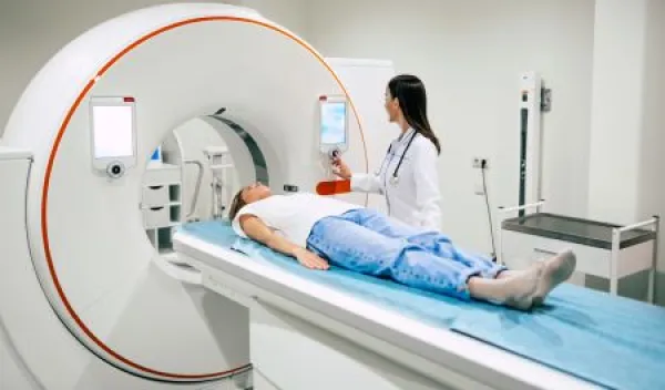
Specialty Service Models & Risk Adjustment, Pinnacle Enterprise Risk Consulting Services