Simplify Mohs Micrographic Surgery Coding
Key concepts every coder must understand. At first glance, Mohs micrographic surgery codes may look like routine excision codes found in the Integumentary section of CPT®. However, coding Mohs surgery requires a deeper understanding of stages, tissue blocks, and the surgeon’s dual role. Even small misunderstandings can lead to significant coding errors and claim denials. In this article, we break down Mohs surgery coding into clear, manageable concepts using CPT® guidance that you can use to code these procedures with confidence. What Is Mohs Micrographic Surgery? According to the CPT® 2026 code book definition, “Mohs micrographic surgery is a technique for the removing of complex or ill-defined skin cancer with histologic examination of 100% of the surgical margins.” A defining feature of Mohs surgery is that the excised tissue is mapped, processed, and examined immediately. This process allows the surgeon to identify and remove additional tissue only where cancer cells remain. This staged approach continues until all margins are clear. Read This Before Coding Mohs surgery coding is fundamentally different from routine excision and destruction procedures, both clinically and from a coding standpoint. Generally, one surgeon performs two roles: According to CPT® guidelines, you should not use the Mohs codes if another provider performs either part of the procedure and reports that part separately. Understand the Staged Nature of Mohs Surgery Routine excisions are typically performed with a single excision in one encounter. In contrast, Mohs surgery is performed in stages during one or more encounters, with each stage involving: Each stage builds upon the previous one until tumor-free margins are achieved. Get to Know the Mohs Codes Mohs surgery codes are reported from 17311-17315, based on anatomic location, stage, and the number of tissue blocks. Primary Mohs Codes (First Stage) Additional Stages (Add-on Codes) These add-on codes are reported in conjunction with the appropriate primary code and represent continued staged excisions. Additional Tissue Blocks This code is reported separately when the surgeon prepares more than five tissue blocks during any stage of Mohs surgery. Remember These Key Pointers to Avoid Mohs Coding Confusion A common area of confusion when using the Mohs codes lies in misunderstanding the difference between stages and tissue blocks. A stage represents a complete cycle of excision and microscopic examination, while a tissue block is an individual segment of the excised tissue prepared for histologic sectioning. Both must be clearly documented to support correct code selection. Another problem using the Mohs codes arises when the anatomic location is misidentified. Correct code selection depends on whether the lesion is located on the head, neck, hands, feet, genitalia, or other complex structures, or the trunk, arms, or legs. Misidentifying the anatomic location can lead to incorrect primary code selection. Know What Is, and What Is Not, Included in the Mohs Codes Routine frozen section staining and the surgeon’s histologic examination by are both included in the codes. This means that you must not report these services separately. However, repairs, flaps, or grafts may be reported separately when supported by documentation. A diagnostic biopsy performed on the same day may be reported separately with modifier 59 (Distinct procedural service) when no prior pathology confirmation exists. Special stains or immunohistochemistry may be reported separately only when non-routine and appropriately documented. Test Yourself With These Coding Examples Scenario 1: Cheek Lesion Mrs. R., a 62-year-old woman, noticed a small, crusted lesion on her left cheek. After evaluation, her dermatologist diagnosed basal cell carcinoma. During Mohs surgery, the dermatologist divided the first stage of tissue removal into four blocks. Microscopic review revealed tumor cells along one margin, prompting a second stage. Three additional blocks were excised, and finally, all margins were clear. The appropriate coding is: Scenario 2: Hand Lesion Mr. K., a 55-year-old construction worker, presented with a firm nodule on the dorsum of his right hand. After evaluation, his dermatologist diagnosed squamous cell carcinoma and performed Mohs surgery. The initial excision yielded six tissue blocks. Microscopic analysis showed remaining carcinoma along one margin. The surgeon performed a second stage with two additional blocks, after which the surgeon confirmed all margins were tumor-free. The appropriate coding is: Know These Key Takeaways Mohs micrographic surgery coding requires careful attention to stages, tissue blocks, anatomic location, and physician roles. By understanding these key concepts, coders can ensure accurate reporting and maintain compliance with CPT® guidelines. Mastery of Mohs coding not only reduces claim denials but also strengthens a coder’s confidence when handling more complex dermatologic procedures. Surbhi Prapanna, CPC-A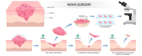
(A version of this article first appeared in the July 2026 issue of AAPC the Magazine)