Neurology & Pain Management Coding:
Know Test Purpose to Differentiate PET, CT, MRI
Published on Tue Oct 06, 2026
Question: Our neurology practice performs positron emission tomographic (PET) scans, as well as CTs and MRIs. What are the differences between the three, for coding purposes?
Nebraska Subscriber
Answer: PET, CT, and MRI are all tests that neurologists perform, albeit for different reasons than physicians in other specialties. The trick is knowing what the test is looking for, how the test is being performed, and the different clinical purposes for each test.
Help is here: During HEALTHCON 2026, Angie Davis CPC, CDEO, ran down the basics for each test during her presentation, “Mastering PET Scan Coding: Keys to Accuracy and Compliance.”
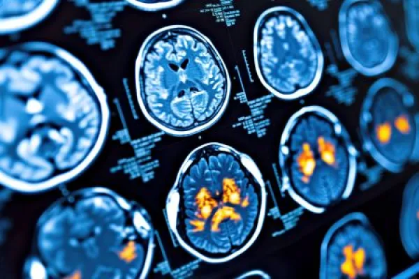
Here’s a look at a list she presented breaking down the difference between PET, CT, and MRI:
- Imaging modality: PET scan
- What it detects: Metabolic and biochemical activity at the cellular level
- Method of detection: Radiopharmaceutical tracer emits positrons detected as gamma photons
- Common clinical uses: Cancer detection and staging, treatment response, cardiac viability, neurologic disorders
- Imaging modality: CT scan
- What it detects: Structural abnormalities and differences in tissue density
- Method of detection: Rotating X-ray beams produce cross-sectional images
- Common clinical uses: Trauma, tumors, fractures, bleeding, organ abnormalities
- Imaging modality: MRI
- What it detects: Soft tissue composition and structural detail
- Method of detection: Strong magnetic field and radiofrequency pulses detect hydrogen proton signals
- Common clinical uses: Brain and spinal cord imaging, ligament injuries, tumors, musculoskeletal disorders.
Chris Boucher, MS, CPC, Senior Development Editor, AAPC