Get Clarity on OCT Coding and Avoid These Pitfalls
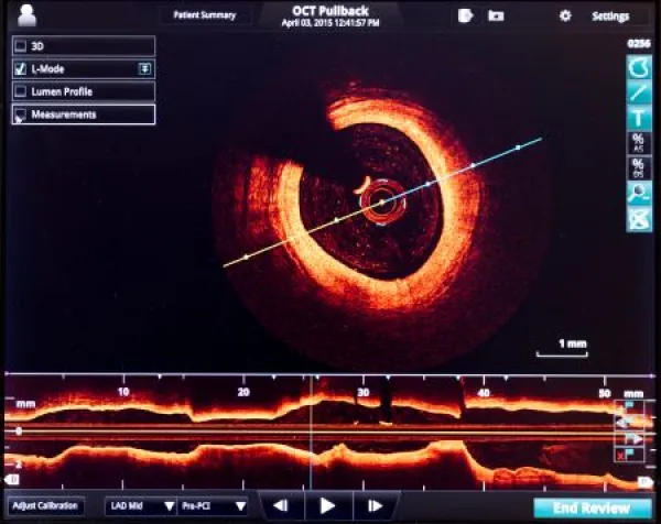
Can you bill OCT separately for each eye? Find out. Since its introduction in the 1990s, optical coherence tomography (OCT) has become a practically routine part of a comprehensive eye exam, especially for patients at high risk for eye disease. One of the most frequently performed diagnostic imaging tests in ophthalmology and optometry, OCT’s ability to provide high-resolution, cross-sectional images of ocular structures, particularly the retina and optic nerve, makes it indispensable for diagnosing, monitoring, and managing a wide range of eye diseases. Because OCT is so widely used, it is also an area of frequent coding errors, denials, and payer scrutiny. Read on to understand what OCT measures, when it is medically necessary, and how to code it correctly for compliant reimbursement. Learn the How and Why of What OCT Evaluates OCT is a noninvasive imaging test that uses low-coherence light waves to generate detailed, cross-sectional images of ocular tissues. The technology is often compared to ultrasound imaging, but instead of sound waves, OCT uses light to achieve micron-level resolution. Clinicians use OCT to visualize and measure retinal layers, optic nerve head anatomy, and nerve fiber thickness — structures that cannot be assessed adequately through routine ophthalmoscopy alone. The test is quick, painless, and does not require contact with the eye, making it suitable for repeated use over time to track disease progression or response to treatment. OCT is most commonly used for diagnosis and longitudinal management, not screening. Its clinical value lies in its ability to detect subtle structural changes before functional vision loss becomes apparent. A provider might use OCT to assess: Other uses include monitoring response to intravitreal injections, assessing unexplained vision loss, and evaluating retinal toxicity from systemic medications. Key point: Payers generally expect OCT to be ordered when results will directly affect clinical decision making, such as initiating, adjusting, or continuing treatment. Keep an Eye on Anterior vs. Posterior Segments Correct coding begins with understanding the distinction between the anterior and posterior sections of the eye as well as the specifics of retinal versus optic nerve OCT. For OCT performed on the anterior (front) segment, report 92132 (Computerized ophthalmic diagnostic imaging (eg, optical coherence tomography [OCT]), anterior segment, with interpretation and report, unilateral or bilateral). A provider would perform anterior OCT to capture detailed images of the front part of the eye, including the cornea, iris, and lens, monitoring for eye conditions such as glaucoma, corneal disorders, and cataracts. To examine the posterior (rear) section of the eye, a provider would look to one of the next three codes in the 9213x series. CPT® code 92133 (Computerized ophthalmic diagnostic imaging (eg, optical coherence tomography [OCT]), posterior segment, with interpretation and report, unilateral or bilateral; optic nerve) describes OTC imaging of the optic nerve head and retinal nerve fiber layer (NFBR). CPT® code 92134 (Computerized ophthalmic diagnostic imaging (eg, optical coherence tomography [OCT]), posterior segment, with interpretation and report, unilateral or bilateral; retina) is used for OCT imaging of the retina, including the macula. Don’t miss: Both code descriptions contain the qualifier “unilateral or bilateral,” meaning that the provider cannot bill separately for each eye. Look to 92137 for OCTA CPT® code 92137 (Computerized ophthalmic diagnostic imaging (eg, optical coherence tomography [OCT]), posterior segment, with interpretation and report, unilateral or bilateral; retina, including OCT angiography), a new code introduced in 2025, describes OCT angiography (OCTA) that does not require an injection of dye like fluorescein angiography 92235 (Fluorescein angiography (includes multiframe imaging) with interpretation and report, unilateral or bilateral) and is noninvasive and infusion-free. Watch out: Codes 92133 and 92134 have long been bundled together as mutually exclusive by the Centers for Medicare & Medicaid Services (CMS) with a “0” modifier, meaning that you cannot report both for the same patient encounter. In 2025, 92137 became bundled as well since the code already includes the OCT portion. Include Both Technical and Professional Services Although some codes are able to be split with modifiers 26 (Professional component) and TC (Technical component …) to bill different portions of the service to different providers, OCT codes include both technical and professional components. This means you have to demonstrate that the provider also performed the interpretation and report — and correct documentation is essential. Medical records should clearly include: Pitfall: Simply stating “OCT performed” or attaching images without interpretation is insufficient and may result in recoupment from payers. Coders should also confirm that the provider performed and documented the interpretation; OCT codes cannot be billed on image acquisition alone. Jerry Salley, BA, MFA, Contributing Writer